Eye Diseases in Pugs
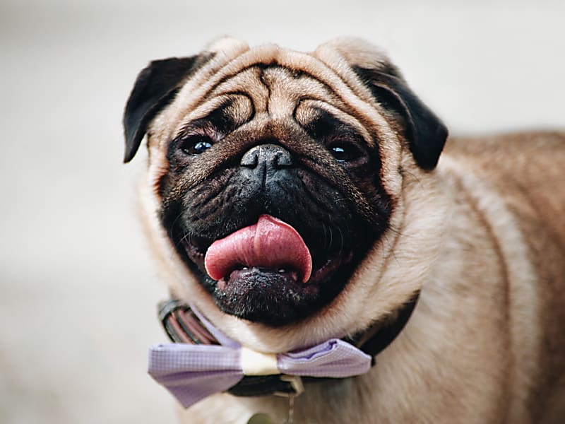
Why are pugs' eyes so big?
In fact, pugs' eyes are not actually larger than those of other dog breeds — rather, their skull is shorter and wider. Because of this, the eye socket is shallower, so the eye sits further forward (physiological exophthalmos) compared to breeds with longer heads.
What eye problems does this skull shape cause?
Pugs' eyeballs are less protected than in longer-headed breeds.
Normally, in dogs, the white part of the eye (sclera) should not be visible. If it is visible, the eyelid is too long (macroblepharon), which makes the eye even more vulnerable.
Because of the short head shape, the folds of skin over the nose turn inward at the inner corner of the eye in pugs (nasal entropion), and the hairs on the eyelid margin continuously irritate the ocular surface. As a result:
- Chronic corneal inflammation develops.
- Pigment deposits form on the eye's surface (pigmentary keratitis).
- This pigmentation reduces vision and can even cause blindness.
What other eye problems are typical for the breed?
Pugs and other brachycephalic (short-headed) breeds (French Bulldog, Shih Tzu, Lhasa Apso) are prone to dry eye disease (keratoconjunctivitis sicca):
- The quantity or quality of the tear film is inadequate.
- The ocular surface doesn't receive enough nourishment and protection.
- Corneal ulcers and inflammation often develop.
- Dry eye disease requires lifelong treatment.
How can I tell if my dog has an eye problem?
- The eyes are often red and discharging.
- Corneal ulcers develop repeatedly.
- The dog sleeps with its eyes open.
- A brownish-grey haze is visible on the eye, especially on the side toward the nose.
- The white of the eye (sclera) is visible.
What solutions exist for these problems?
The eyelid positioning abnormality can be corrected surgically (nasal canthoplasty):
- Both the lower and upper eyelids are shortened.
- The eyelid is turned outward so that the hairs no longer irritate the ocular surface.
In addition, pugs usually require lifelong ophthalmic treatment:
- Eye drops containing cyclosporine, pimecrolimus, or tacrolimus.
- These help remove pigment that has already deposited and prevent further pigment formation.
How does the surgery work?
The surgery is performed under general anesthesia. Anesthesia:
- Always carries some risk, although this is minimal in healthy animals.
- Due to the short head shape, the airways are narrower, which slightly increases the risk.
- The procedure is carried out using anesthetic gas, delivered to the lungs through an endotracheal tube.
- During the recovery phase, the patient is kept under continuous monitoring.
What home care is needed after the surgery?
For 2 weeks after the surgery:
- The dog must wear an Elizabethan collar.
- Eye ointment must be applied 3 times daily.
- Pain relief must be given once daily.
- Suture removal takes place 10–14 days after the surgery.